
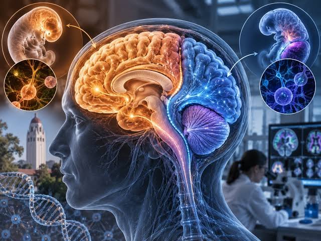

A new Stanford Medicine study is challenging the traditional view of the human brain as a single, unified organ.
Researchers found evidence that the front and back of the brain develop from different groups of progenitor cells during embryonic development.
The findings were published in Nature Neuroscience and could change how scientists understand brain development and neurological diseases.
Front and Back of Brain Have Different Origins
The researchers identified two distinct progenitor cell populations involved in brain development.
One population expresses the Otx2 gene. It develops into the forebrain and midbrain.
These areas are involved in functions such as language, consciousness and abstract reasoning.
The second population expresses the Gbx2 gene. It develops into the hindbrain.
The hindbrain controls many essential automatic functions. These include breathing, heartbeat and sleep.
Senior study author Kyle Loh said the research provides evidence that the front of the brain develops from a completely different progenitor cell population than the back.
Cells Follow Separate Developmental Paths
The researchers found that the two cell populations do not overlap during development.
They also have different chromatin structures.
Chromatin helps organize DNA and control which genes are active inside cells.
According to the researchers, these differences keep the two populations on separate developmental pathways.
This discovery may help explain a long-standing problem in neuroscience.
Scientists have struggled to reliably produce hindbrain neurons from human stem cells in laboratory settings.
The new understanding of their developmental origins has now provided a potential solution.
Scientists Create Hindbrain Motor Neurons
Using the newly identified developmental pathway, researchers successfully converted human pluripotent stem cells into functional hindbrain motor neurons.
This could provide scientists with a new laboratory model for studying diseases that damage these neurons.
Among them are spinal muscular atrophy (SMA) and amyotrophic lateral sclerosis (ALS).
Both conditions can affect motor neurons and may interfere with important functions such as swallowing and breathing.
Researchers could potentially use laboratory-grown neurons to better understand how these diseases develop.
The cells could also help scientists investigate possible treatments and regenerative therapies.
Evolutionary Origins May Be Hundreds of Millions of Years Old
The researchers found a similar developmental pattern in several other species.
The pattern was observed in chickens and zebrafish.
It was also identified in acorn worms, an organism belonging to a much earlier branch of animal evolution.
This suggests that the developmental division may have ancient evolutionary origins.
Researchers estimate that the pattern could date back at least 550 million years.
The finding indicates that the separation between the developmental systems responsible for different brain regions may have emerged very early in animal evolution.
Potential Impact on Neurological Research
The study could have implications beyond understanding how the brain develops.
Being able to produce specific types of human neurons in the laboratory could give researchers better tools for studying neurological disorders.
It may also support future research into regenerative medicine.
However, the findings represent an advance in understanding brain development rather than an immediate treatment for neurological diseases.
Researchers will need further studies to determine how the discovery can translate into therapies.
The study nevertheless provides new evidence that the brain’s complex structure may have developed through distinct cellular pathways rather than from one uniform developmental system.